Heart Dissection
Aim
The aim of this experiment was to successfully dissect a heart and identify all of the chambers and parts of it.
Appearance & texture
The heart had two different colours: pale pink (fat) and a reddy-brown. You could see where all of the arteries and veins begun. You could also see all of the blood vessels on the surface of the heart.
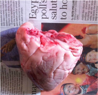
An outside view of the heart before we made any incisions.
Blood Vessels
On the surface of the heart muscle, there are coronary arteries. They carry nutrients and oxygen to the heart muscle. They are a darker colour than the rest of the heart and the kind of sink in to the surface.It these arteries were blocked by a clot, I think it would restrict the amour of blood flowing to the rest of the body and you would not get enough nutrients or oxygen.

A top view of the heart showing where all the blood vessels enter and exit.
Four Chambers
The heart is made up of 4 chambers: the left atrium, right atrium, left ventricle and right ventricle. The left ventricles muscle wall is the thickest out of all four chambers. It is also a lot stronger.
The thickness of the muscles at the top of the heart had quite a strong, boney feel. The muscles at the bottom of the heart were softer and flexible but they were a lot thicker. Surrounding the heart is a layer of fat. There was a lot more fat covering the top half of it. Also, at the top of the heart, there are 4 holes where the major blood vessels enter. The muscle wall thickness changes depending on which blood vessel it is.
The thickness of the muscles at the top of the heart had quite a strong, boney feel. The muscles at the bottom of the heart were softer and flexible but they were a lot thicker. Surrounding the heart is a layer of fat. There was a lot more fat covering the top half of it. Also, at the top of the heart, there are 4 holes where the major blood vessels enter. The muscle wall thickness changes depending on which blood vessel it is.


Pulmonary Artery
Deoxygenated blood leaves the right ventricle in an artery and travels to the lungs. Here, the blood collects oxygen so it is now oxygenated. The blood travels back to the heart via the vein.
Aorta
The aorta is an artery with a very thick muscle wall. It has to be thick so it can pump hard enough for the blood to travel around the body. The aorta takes oxygenated blood to the brain and the rest of the body. The aorta was quite hard to cut because of how thick its wall was. It had a slimy, squishy feel yet you could tell it was strong.
Vena Cava
The vena cava is the vein that takes deoxygenated blood back to the heart. It has a thinner muscle wall compared to the aorta as it doesn't have to deal with such a high blood pressure. The vena cava goes back into the right atrium. When we poured the water into the vena cava, it went through the heart and came out of the pulmonary artery.
Pulmonary Vein
When we poured the water into the pulmonary vein, it came out through the aorta.
Left Chambers.
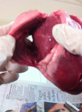
When you cut into the left side of the heart and look at the left ventricle, you can see the thick muscle wall. You can also the valves separating the two left chambers. The valves job is to make sure the blood flows in one direction and doesn't go back to where is has already been.

The left ventricle.
Right Chambers
Both of the right chambers were a lot smaller than the left. Its muscle wall was a lot thinner but you could see the valves quite clearly.
The right ventricle.
Parts and Functions
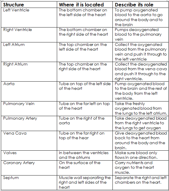
The table below displays information about the different parts of the heart, where they are located and what they do.
Comparing and Analysing
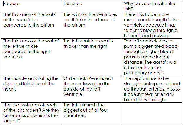
Below is a table that shows comparisons and analysis' of different parts of the heart.
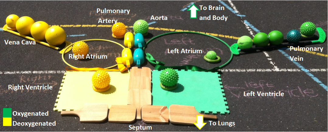
An abstract representation of a heart showing the way that the oxygenated and deoxygenated blood travels.
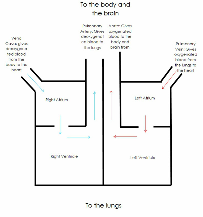
This is a box heart diagram showing the direction of the blood flow.
